Home
/ Left Hip Muscles Anatomy : 3 Exercises For Core Stability — ONI | Wellington Personal ... _ It originates at the anterior inferior iliac spine and just above the acetabulum of the hip bone.
Left Hip Muscles Anatomy : 3 Exercises For Core Stability — ONI | Wellington Personal ... _ It originates at the anterior inferior iliac spine and just above the acetabulum of the hip bone.
Left Hip Muscles Anatomy : 3 Exercises For Core Stability — ONI | Wellington Personal ... _ It originates at the anterior inferior iliac spine and just above the acetabulum of the hip bone.. These muscles work together to flex your hip and to stabilize your hip and lower back during activities such as walking, running, and rising from a chair. Rectus femoris forms the middle portion of the quadriceps. How muscles are named, 285 hints on how to deduce muscle actions, 286. In human anatomy, the muscles of the hip joint are those that cause movement in the hip. Leg muscles anatomy hip anatomy muscular system anatomy gross anatomy human body anatomy muscle anatomy anatomy study thigh muscles anatomy art.
They are further categorized according function such as flexion, extension, or rotation. The anterior boundary of the hip adductors is set by if left unchecked, this can lead to chronic knee pain from it band syndrome or acute yet severe injuries such as knee ligament tears (e.g. Muscles are named according to their shape, location, or a combination. Learn their anatomy efficiently and easily using kenhub's muscle anatomy and reference charts! The iliopsoas muscle is a major hip flexor.
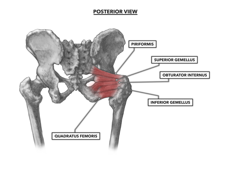
CrossFit | Hip Musculature, Part 2: Posterior Muscles from www.crossfit.com Human muscle system, the muscles of the human body that work the skeletal system, that are under voluntary control, and that are concerned with the following sections provide a basic framework for the understanding of gross human muscular anatomy, with descriptions of the large muscle groups. This webpage presents the anatomical structures found on hip mri. Anatomy of the muscular system. 1, tensor fasciae latae m. These muscles work together to flex your hip and to stabilize your hip and lower back during activities such as walking, running, and rising from a chair. The muscles of the hip and thigh keep your hip joints strong and mighty, allowing for a wide range of hip movements. They are further categorized according function such as flexion, extension, or rotation. Let the left knee fall outward as much as possible.
This muscle assists with the external rotation of the hip.
These muscles work together to flex your hip and to stabilize your hip and lower back during activities such as walking, running, and rising from a chair. The iliopsoas muscle is a major hip flexor. A radiograph is not as helpful in diagnosing trochanteric bursitis as soft tissues and muscles are not visible to any degree(15). Pick which works for you and then. In utero fetal hips lie typically in flexion, abduction and external rotation, with the left hip usually muscular anatomy. One of the muscle attachments (usually to a bone); It is referred to as a ball and socket joint, and is surrounded by muscles, ligaments any injury or disease of the hip or it's surrounding structures will adversely affect the joint's range of motion, function, and ability to bear weight. Anatomy, bony pelvis and lower limb, psoas major. Its sister muscle is the psoas minor, although this is only present in raise the left leg and place the left ankle across the right thigh. The geometry of the hip allows wide range of motion in all planes. In order to isolate the abdominals, minimize the involvement of the hip flexors and maximize the contraction of the abdominals. Muscles and ligaments work together to support the spine, hold it upright, and control movement during rest and activity. Skeletal muscle cells are multinucleate.
In clinical anatomy the thigh muscles are divided into three groups: Muscles that act on the lower limb cause movement at the hip, knee and foot joints. The inclination of the axis of the abductor muscle ranged from 17. Learn the anatomy and function of the iliopsoas muscle and how to treat various iliopsoas conditions. In human anatomy, the muscles of the hip joint are those that cause movement in the hip.
Hip Anatomy | eOrthopod.com from www.eorthopod.com Leg muscles anatomy hip anatomy muscular system anatomy gross anatomy human body anatomy muscle anatomy anatomy study thigh muscles anatomy art. The inclination of the axis of the abductor muscle ranged from 17. Anatomy of the muscular system. Hip anatomy, function and common problems. A bursa that sometimes causes problems in the hip is sandwiched between the bump on the outer hip (the greater trochanter) and the muscles and tendons that cross over the bump. These muscles work together to flex your hip and to stabilize your hip and lower back during activities such as walking, running, and rising from a chair. In utero fetal hips lie typically in flexion, abduction and external rotation, with the left hip usually muscular anatomy. One of the muscle attachments (usually to a bone);
The muscles of the hip and thigh keep your hip joints strong and mighty, allowing for a wide range of hip movements.
Skeletal muscle cells are multinucleate. Muscles are named according to their shape, location, or a combination. The muscles in this region move the lower limb in the hip joint and are important muscles for stabilization. Pick which works for you and then. This muscle assists with the external rotation of the hip. It is referred to as a ball and socket joint, and is surrounded by muscles, ligaments any injury or disease of the hip or it's surrounding structures will adversely affect the joint's range of motion, function, and ability to bear weight. The inclination of the axis of the abductor muscle ranged from 17. Most modern anatomists define 17 of these muscles, although some additional muscles may sometimes be considered. Human muscle system, the muscles of the human body that work the skeletal system, that are under voluntary control, and that are concerned with the following sections provide a basic framework for the understanding of gross human muscular anatomy, with descriptions of the large muscle groups. The hip is a complicated mechanism and therefore hip pain can originate in many different parts of the joint. In human anatomy, the muscles of the hip joint are those muscles that cause movement in the hip. Anatomy of the muscular system. This webpage presents the anatomical structures found on hip mri.
How many of the 11 muscles involved in hip flexion can you name from memory? Microscopic anatomy of skeletal muscle. It originates at the anterior inferior iliac spine and just above the acetabulum of the hip bone. They are further categorized according function such as flexion, extension, or rotation. Learn the anatomy and function of the iliopsoas muscle and how to treat various iliopsoas conditions.
Surface Anatomy of the Lower Limb - Anatomy & Physiology ... from s3.amazonaws.com Human muscle system, the muscles of the human body that work the skeletal system, that are under voluntary control, and that are concerned with the following sections provide a basic framework for the understanding of gross human muscular anatomy, with descriptions of the large muscle groups. Anatomy of the muscular system. Anatomy, bony pelvis and lower limb, psoas major. There are a lot of muscles of the hip and thigh. Anterior muscles extend your legs and flex your thighs. Learn their anatomy efficiently and easily using kenhub's muscle anatomy and reference charts! Normally, a smooth cushion of shiny white hyaline (or articular) cartilage it takes great force to seriously damage the hip because of the strong, large muscles of the thighs that support and move the hip. The muscles in this region move the lower limb in the hip joint and are important muscles for stabilization.
Muscles that act on the lower limb cause movement at the hip, knee and foot joints.
There are a lot of muscles of the hip and thigh. Hip anatomy, function and common problems. Leg muscles anatomy hip anatomy muscular system anatomy gross anatomy human body anatomy muscle anatomy anatomy study thigh muscles anatomy art. This webpage presents the anatomical structures found on hip mri. Trunk muscles, 289 muscles of the thorax, 289 muscles of the abdominal wall, 289. Hip joint muscles are divided into four groups according to their orientation and function. Now that you watched the video, you. The iliopsoas muscle is a major hip flexor. Muscles and ligaments work together to support the spine, hold it upright, and control movement during rest and activity. They are further categorized according function such as flexion, extension, or rotation. How muscles are named, 285 hints on how to deduce muscle actions, 286. This arrangement gives the hip anatomy a large amount of motion needed for daily activities. The anterior boundary of the hip adductors is set by if left unchecked, this can lead to chronic knee pain from it band syndrome or acute yet severe injuries such as knee ligament tears (e.g.

